Keywords:
Neuroradiology brain, CT, CT-Angiography, Contrast agent-intravenous, Computer Applications-Detection, diagnosis, Haemorrhage
Authors:
Y. Watanabe, H. Tanaka, M. Nishizawa, Y. Kunitomi, A. Tsukabe, N. Tomiyama; Suita/JP
DOI:
10.1594/ecr2012/B-0503
Methods and Materials
Subjects
• Twenty-six ER patients with intracranial hemorrhage who had underwent dual-energy CTA were retrospectively included in this study. (Male:Female=16:10,
Mean age 60-year-old,
16-84 year-old).
Patients consists of idiopathic intracerebral hemorrhage (ICH) 12pts,
traumatic hemorrhage 11 pts,
subarachnoid hemorrhage + ICH 2 pts,
and intra-tumor hemorrhage 1 pts.
Methods
• All patients had undergone dual-source dual-energy CTA (100kV and 140kV) and delayed-phase enhanced CT (2 minutes after contrast injection) between April 2010 and July 2011.
• CT was performed with a dual-source CT unit (Somatom Definition Flash,
Siemens Healthcare) operated in the dual-energy mode,
with tube A at 100kV and tube B at 140kV 100 and 140 kV.
• Dual-energy CT images were post-processed with commercial software applying a three-material-decomposition algorithm (materials: iodine,
hematoma,
water) to calculate iodine images and virtual unenhanced images and also to generate combined CT images that create the impression of 120 kV CT images.
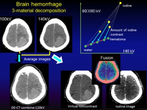
Fig. 1: Diagram of image postprocessing of DE-CT
Image analysis
• Two neuroradiologist,
blinded to the patient’s data,
reviewed the combined images first,
and then the iodine images with combined images to detect contrast enhancement or contrast leakage in the hematoma,
with final decisions made by consensus.
• The contrast enhancement of delayed CT is defined as the reference standard.
